Clubfoot. Diagnosis and clinical evaluation
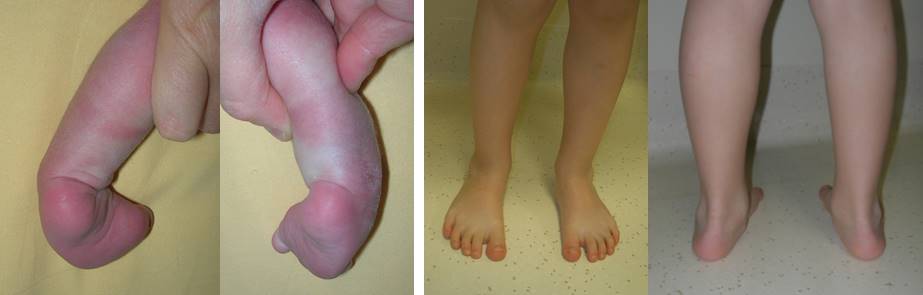
Congenital clubfoot is one of the most frequent deformities in pediatric orthopedics (1-2 newborns in 1000, mainly males). It consists of a complex, three-dimensional deformity of the foot that cannot be reduced (that is, the position cannot be completely corrected by passive manipulations).
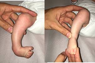
The deformity of clubfoot can be subdivided into three main features: equinus (the foot points down and the ankle can not be moved in dorsal flexion, that is, upwards), varus (the heel, seen from behind, is deviated inward) and adductus (the foot, seen from below, is deviated inwards).
What are the causes of clubfoot?
Causes, in most cases, are unknown (idiopathic clubfoot): environmental and genetic factors play a role (multifactorial origin).
The deformity develops in the first months of pregnancy (around 3 months) and is not due to a uterine compression of the foot in the last period of pregnancy. In a high percentage of cases, the diagnosis of clubfoot can be performed already during the ultrasound examination performed during the second trimester.
Less frequently, clubfoot is associated with other problems (secondary clubfoot): myelomeningocele, arthrogriposis, hypoplasia, neuromuscular pathologies, amniotic band syndrome, various syndromic diseases, etc.
Initial assessment
At initial assessment, the clubfoot is differentiated from normal conditions: in some cases a diagnosis of clubfoot is made during pregnancy and then excluded at birth (false positive).
Then, true clubfeet are differentiated from positional foot deformities. These deformities can seem quite similar: yet, in congenital clubfeet the deformity develops in the first period of pregnancy and is fixed (not completely reducible), in positional deformities the foot has a normal development but is pushed into deformity during the last period of pregnancy and this deformity is reducible. Treatment and prognosis of a simple positional foot deformity are less complex.
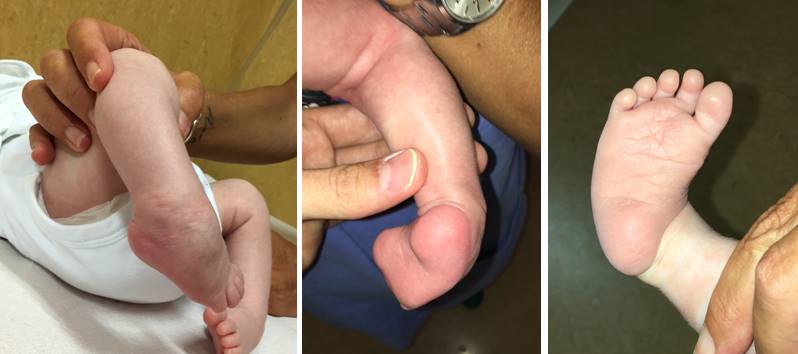
Severity score
The severity of the deformity is evaluated at the initial assessment. Some internationally recognized scores are used, for example the Pirani score ranges from 0 to 6 points (0 no deformity, 6 severe deformity, and the Dimeglio score from 0 to 20 points (20=severe deformity). These scores consider the possibility to reduce the deformity by manual manoeuvres and some specific morphological aspects (for example, creases).
Other important features, such as stiffness and muscle function, are also considered.
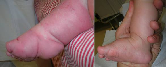
Atypical clubfoot
Atypical clubfoot is characterized by a short and rigid foot, often with deep skin folds. These feet are more difficult to treat, generally require a higher number of casts and have a higher tendency of cast slipping. Clubfeet can become atypical as a result of an initial wrong treatment.
The treatment. Ponseti method.
The Ponseti method is the most widely used method for the treatment of congenital clubfoot. It was conceived by Dr. Ignacio Ponseti in the ’60s. The deformity is corrected progressively in order to limit the use of invasive surgery.
The treatment consists of three different phases.
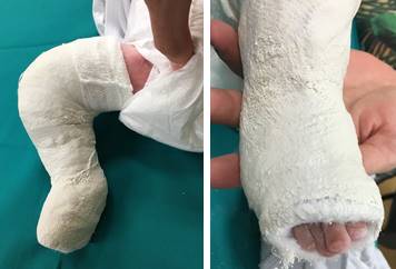
1- CORRECTIVE CASTS.
The correction is gradual, through weekly sessions. At each session, the cast is removed, corrective manipulations are performed, and a cast is applied in the desired position. Manipulations are not painful or aggressive. Casts should be long from the thigh to the toes, with the knee flexed at a right angle. Toes must be visible enough to be controlled.
During the days spent in the cast, the structures of the foot (bones, joints, ligaments, etc.) relax, and are ready to be manipulated in a more corrected position at the successive session.
The number of casts needed depends on many factors, including rigidity and severity: 2 to 3 casts are sufficient for less severe cases, up to six – seven casts are required for more severe feet.
2- PERCUTANEOUS TENOTOMY OF THE ACHILLES TENDON.
During the first phase, the foot is progressively rotated outwards, but can not be corrected in dorsal flexion due to the shortening of the Achilles tendon. This feature can not be corrected by manipulations.
Once the correction of the other deformities has been done, a percutaneous tenotomy of the Achilles tendon is performed. This is a small surgical procedure, in which the Achilles tendon is completely sectioned percutaneously (no incision is made to visualize the tendon), thus eliminating the final obstacle to equinus correction. The tendon heals within a period of 2 to 3 weeks, and the incision typically leaves only a small scar of 2-3 mm.

Upon completion of this stage, dorsal flexion of the ankle can be performed without risk of deforming the bones, thus completing the correction of the clubfoot. A last cast is applied for 15-20 days.
The procedure is usually performed under local anesthesia.
Tenotomy is required in about 70-80% of clubfeet. In some milder cases, a spontaneous progressive relaxation of the posterior structures (Achilles tendon, triceps, etc.) occurs during the first phase, so that the tenotomy is not required.
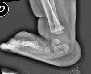
The decision to perform the tenotomy is taken only when the other deformities are corrected. If there is still some doubt, an X-ray could be taken to directly visualize the relationship between the various bones.
3- MAINTENANCE OF CORRECTION.
Once the last cast has been removed, the foot is completely corrected, but has the tendency to recur.
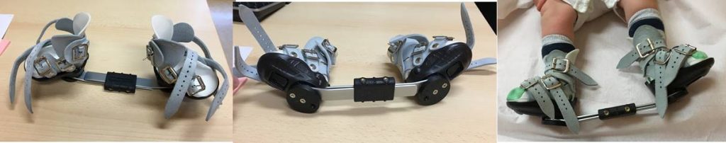
The most effective way to maintain the correction achieved and to prevent recurrence is the use of a brace, called “abduction brace”.
The abduction brace consists of two shoes joined together by a bar, which keep the feet rotated outwards. The same brace is also used when only one foot is affected. In this case, the non-pathological foot is kept less rotated outwards than the affected foot. In the case of bilateral clubfoot, the rotation is usually 60-60 degrees, and in the case of unilateral clubfoot, the rotation is usually 30-70 degrees. Sometimes the doctor will decide to change the rotation according to the foot’s characteristics.
This brace is generally well tolerated (families report a few days necessary to get used to). The knees and hips are free to move.
How long should the brace be worn?
The brace should be worn:
- 23 hours per day for the first 3 months.
- Then, 18-19 hours with the brace (5-6 hours without brace) are suggested for one month.
- Then, a progressive reduction of use is suggested (one hour of reduction each month) until 13-14 hours of use
- When the child begins to walk, the brace should be worn for about 12 hours, corresponding to night and nap time.
Since the risk of recurrence is higher in the first years of growth and then decreases, it is generally recommended to use the brace at least until the age of five years.
What are the outcomes of the Ponseti method?
When performed by a properly trained doctor, the Ponseti method allows the correction of idiopathic clubfeet in over 95% of cases.
A properly treated clubfoot generally does not interfere with normal life, including sports participation and the use of normal shoes.
In secondary club feet, particularly in cases associated with general stiffness (for example arthrogryposis), the percentage of correction is lower.
Once the correction has been obtained, follow up visits are needed until skeletal maturity. The main causes of recurrence of the clubfoot are: the non-use of the abduction brace, the lack of complete correction at the initial treatment and the imbalance of muscle function. Relapse can affect any aspect of the deformity (equinus, varus, cavus, etc.) and must be recognized and treated promptly.
The recommended treatment in case of relapse in the first years of life is recasting, which means repeating manipulations and corrective casts; if, at the end of this treatment, the deformity is corrected, the brace is resumed. If a limited dorsiflexion persists (recurrence of equinus), it may be indicated to repeat the percutaneous tenotomy of the Achilles tendon.
Children over 3-4 years of age who have a recurred clubfoot can be treated with a series of corrective casts associated with the tibialis anterior tendon transfer.
Why choose OrtoPediatria for clubfoot treatment?
Properly trained doctors are needed to get the higher success rate. Also in follow up visits, experience is essential for early detection of a possible relapse.
OrtoPediatria is a team of paediatric orthopaedic surgeons specialized in the treatment of congenital clubfoot with the Ponseti method, through their experience in Italy and abroad.
A group of doctors dedicated to this pathology guarantees the patient a high level of competence and efficiency.
You might be interested to:
Back to the Clubfoot – Center of Excellence