Clubfoot relapse: tibialis anterior transfer and other surgical procedures
Although the deformity of the clubfoot has been successfully corrected in the first months of life, there is a significant risk of relapse, that is, a risk that the deformity may recur, partially (that is, only in one of its components, for example equinus) or totally (equinus-varus-adductus).
This risk is influenced by several factors.:
- The initial treatment of the foot was not adequate, and the slight residual deformity slowly led to a relapse of the foot.;
- Failure to use the abduction brace: it represents the most frequent cause of relapse in the first years of life during treatment with the Ponseti method;
- True recurrence: even a perfectly corrected foot can recur, due to the underlying problem (that is, the same cause that caused the club-foot at birth). Several studies are underway to clarify these aspects, but probably the problem is related to the altered growth of the postero-medial musculature that does not develop at the same rate as the remaining muscles.;
- Underlying pathology: in case of injured feet relapsed, especially when there are multiple relapses, it is necessary to suspect the role of an underlying neuromuscular pathology that impairs the correct development of the feet;
- Muscle imbalance: if there is a prevalence of the muscles that carry the foot inward compared to the muscles that carry it outward, the foot may tend to relapse.;
- Scar retractions: in the case of feet with long scars and adherences between the different tissues, due to previous surgical treatments.
The relapse needs to be recognized promptly. Due to the increased risk of recurrence, all patients treated for congenital clubfoot should carry out periodic checks during growth. It is essential to evaluate foot assessments correctly, by describing the various aspects of the foot (e.g., the degrees of dorsiflexion) in order to understand if the deformity is stable or if it is worsening.
When a relapse is recognized, treatment must be started immediately, without waiting for further worsening that would require more invasive treatment.
The Ponseti method for treatment of clubfoot relapses
Younger children usually respond well to the Ponseti method when there is a relapse.
Handling and packaging of progressive corrective cast devices are carried out during sessions (so-called “recasting”).
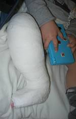
At the end, if there is still residual equinus, lengthening of the percutaneous Achilles tendon is done.
The application of plasters and braces is the main problem.
To apply a plaster, it is important to use tricks, such as video games, to keep him quiet and distracted. Usually, you don’t have to do this with sedation.
Younger children are more resistant to accepting the brace than neonatal infants, and parental involvement is critical for success. For cases that relapse due to poor use, it is necessary for the family to become aware of the braces’ usefulness.
With this method, it is possible to achieve results even with feet that were initially treated with other methods. In the case of feet initially treated with surgery, the Ponseti method can still lead to correction, avoiding the need for additional surgery. This avoids the creation of additional adhesions, scars, and fibrosis.
Tibialis anterior transfer for treatment of clubfoot recurrence
In older children over 3 years of age, cases of recurrence of deformities are usually treated with a series of corrective plasters. Then, a procedure is used to prevent the deformity from coming back. This procedure is called a lateral transfer of the anterior tibial muscle.
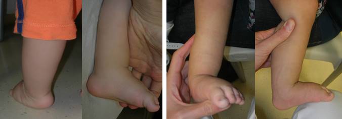
The tibialis anterior is a muscle that allows active dorsal flexion of the foot. In other words, it allows you to lift the foot upwards while walking. It inserts on the first cuneiform bone and the base of the first metatarsal bone, which are on the dorsal and internal part of the foot.
The tendon of the tibialis anterior muscle can be moved to a more lateral position by detaching it from its original position and reattaching it to the third cuneiform bone. In this way, at each step, the muscle pulls the foot more outwards.
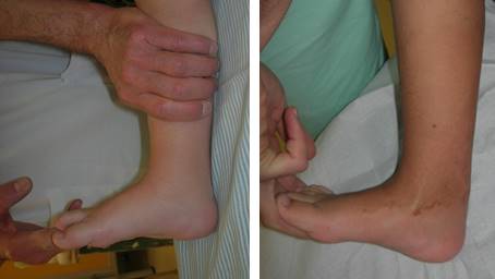
This technique is widely used, both in cases of marked and persistent dynamic supination (feet that are well corrected, but walk shows a marked tendency to supination), and in relapsed feet (usually preceded by a series of corrective plasters).
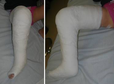
There are several surgical technique options: two or three incisions, passing the tendon over or under the retinal, etc. One point of discussion is how the tendon is fixed at the point where it is re-inserted. The traditional technique involves passing the tendon through a bone tunnel and fixing it under the sole of the foot with sutures (possibly using a button). This technique requires a double period of immobilization. The first period is with a plaster without weight-bearing, which allows the tendon to connect with the bone. The second period is with a plaster that allows weight-bearing. More recent techniques involve the use of metal screws or anchors to fix the tendon to the bone. This form of fixation is immediately effective and allows the patient to walk earlier, which speeds up the functional recovery. However, the possible risk of failure of the screw (detachment of the screw from the bone) must be considered.
The results of this procedure are satisfactory, and the improvements will be evident over time, as a result of the dynamic action of the transferred tendon.Unfortunately, there are cases where the feet recur even after the anterior tibial has been transposed, so it is important not to lower your guard even after the procedure and throughout growth.
Other problems and possible surgery for clubfoot relapse
The risk of clubfoot relapse is significant until the age of 4–5 years (mainly between 2 and 4 years) and then decreases. It is advisable to carry out periodic checks until the skeletal maturity because the risk does not disappear completely during growth.
However, this is especially true for cases where the feet were treated correctly and completely at birth.
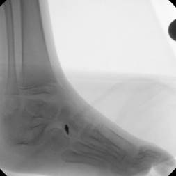
In most cases, the consequences of incomplete corrections, or articular alterations are highlighted in growth cases.
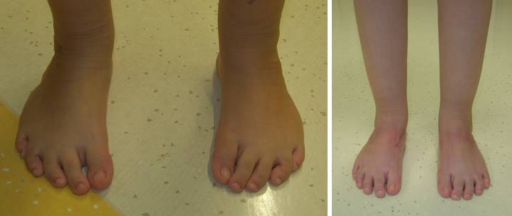
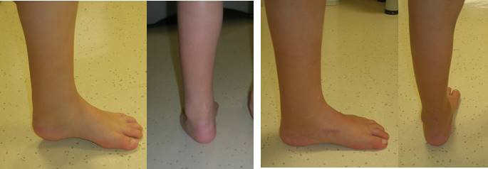
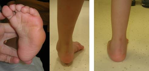
Among the possible problems, in addition to the recurrence of deformities (equinus, varus, adduction, supination, cavism), we mention:
– subluxation of the scaphoid
– flattening of the talus
– joints’ stiffness
– bone deformation
– injuries to growth cartilage
– hypercorrection, with the foot drooping in valgus-pronation or talus
– dorsal Bunion (dorsal flexion of the 1 metatarsal and plantar flexion of the big toe)
Such alterations must be recognized, and the treatment must be directed to the specific problem present.
The principles of treatment are the same as the Ponseti method, that is, try to minimize the surgical aggression and preserving as much as possible the joints.
However, in older patients, the deformities are often so severe that surgery must also include osteotomies in addition to possible soft tissue releases.
Here is a short list of some procedures that may be needed:
- Percutaneous lenghtening of the Achilles tendon and percutaneous plantar fascia release
- Plantar fascia release: the insertion of the plantar band is detached (for residual cavus)
- Shortening osteotomies of the lateral column (of the calcaneum or cuboid)
- Medial and/or posterior soft tissue release: elongation of the medial and posterior soft tissue in cases where it is not sufficient to carry out percutaneous procedures
- Corrective calcaneal osteotomies (varus or valgus)
- Osteotomies of the base of the first metatarsal: to raise or lower a first metatarsal when it points too downwards or upwards
- Arthrodesis: that is, fusions of the joints. To be carried out only at the end of the growth of the foot and in cases of rigid or arthrosic joints
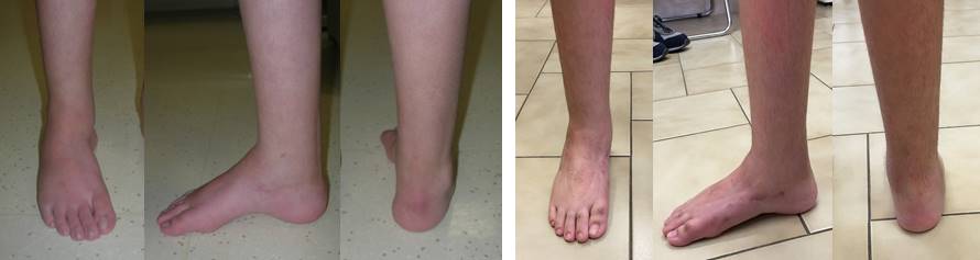
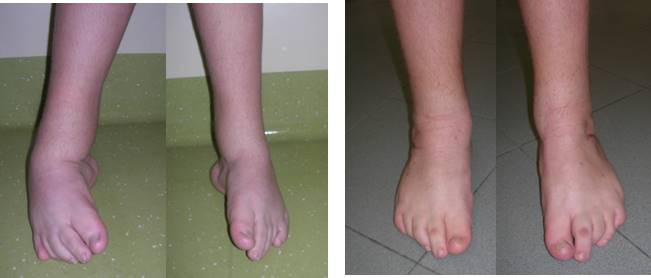
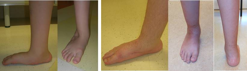
Here are some examples.
In case of a clubfoot relapse, why contact the OrtoPediatria?
It’s important to treat clubfoot early so that the situation doesn’t get so bad that it needs aggressive treatment.
As highlighted in this article, Ortopediatria has considerable experience in the treatment of clubfoot relapses, using the most modern principles of the Ponseti method.
You might be interested to:
Back to the Clubfoot – Center of Excellence