What is congenital pseudoarthrosis of the clavicle?
Congenital pseudoarthrosis of the clavicle is a very rare congenital abnormality, characterized by a lack of fusion of the medial and lateral ossification centers of the clavicle.
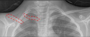
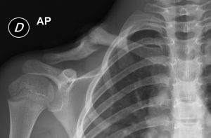
The clavicle shows an interruption in its central portion and the bone is divided into two portions, one lateral and the other medial.
The exact causes are still not fully known. It is likely the result of damage to the ossification process of the clavicle during the fetal phase (probably due to direct compression by the right subclavian artery).
The deformity is predominantly unilateral, almost always in the right side; it occurs exceptionally in the left side (especially patients with congenital inversion of the viscera, called “situs inversus”). In 10% of cases the presentation is bilateral.
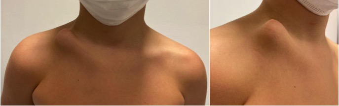
Children with congenital pseudoarthrosis of the clavicle sometimes show a local, painless swelling at the site of pseudoarthrosis in neonatal age. This condition can be confused with a fracture of the clavicle at birth. The main difference is the absence of pain and functional impotence (typical of a fracture).
In most cases, the diagnosis of congenital pseudoarthrosis of the clavicle is delayed, when the deformity becomes more evident with growth and asymmetry of the shoulder girdle appears.
In some cases it can be associated with pain in the shoulder and limitation of the range of movement. Rarely neurovascular disturbances occur.
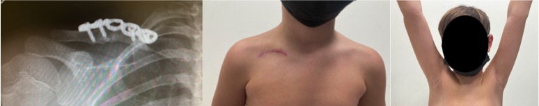
The diagnosis is confirmed by the radiographic examination, which shows a characteristic picture of a clear separation of the central part. The medial fragment is typically higher than the lateral one, being pulled by muscular forces.
Treatment of congenital pseudoarthrosis of the clavicle.
Treatment is still much discussed.
• In the absence of symptoms and in cases with scarce aesthetic disturbances, surgical treatment is not recommended and follow up of the deformity is recommended.
• In children with symptoms (pain, etc) or functional limitations, and in patients with very obvious cosmetic defect, surgery is recommended.
Surgery for the treatment of congenital pseudoarthrosis of the clavicle is usually recommended between 5 and 9 years.
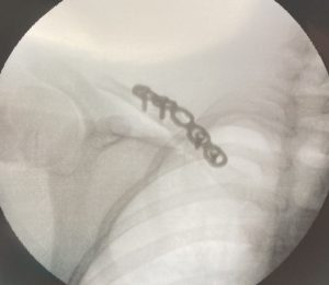
The procedure includes excision of the site of pseudoarthrosis, regularization of the two stumps (with removal of the pathological bone), opening of the medullary canal, application of bone grafts (usually taken from the iliac bone) and stabilization with plate and screws.
In the past, stabilization was carried out with other systems, such as Kirschner wires or intramedullary nails, followed by a long immobilization in bandage (Desault) or cast (toracobrachial).
Currently these stabilization techniques are reserved only for smaller patients and have been replaced by the use of plates and screws. Plates generally provide greater rate and speed of healing, greater stability, and allow shorter and lighter immobilization (brace).
Results of surgical treatment.
The results of surgical treatment are very good, with functional improvement of shoulder movement and aesthetic improvement.
Main possible complications are the lack of consolidation at the site of pseudoarthrosis, infection and wound problems, and are largely dependent on the surgical technique used.
Several studies have found a greater incidence of lack of consolidation and infection when stabilization with Kirschner wires or intramedullary nails was used; wound problems are more frequent when plates and screws are used.
The right surgical indication (choice of the patient, surgical planning), the control of possible complications and an adequate rehabilitation are certainly the basis of greater results in the treatment of congenital pseudoarthrosis of the clavicle.
BIBLIOGRAPHY
- https://www.orthobullets.com/pediatrics/4039/congenital-pseudoarthrosis-of-clavicle
- Kim – Congenital pseudarthrosis of the clavicle surgical decision making and outcomes – JSES 2019
- Chandran – Congenital clavicular pseudarthrosis. Comparison of two treatment methods – JCO 2011
- Studer – Operative treatment of congenital pseudarthrosis of the clavicle a single-centre experience – JPO 2016
Back to Pediatric Orthopedics